-
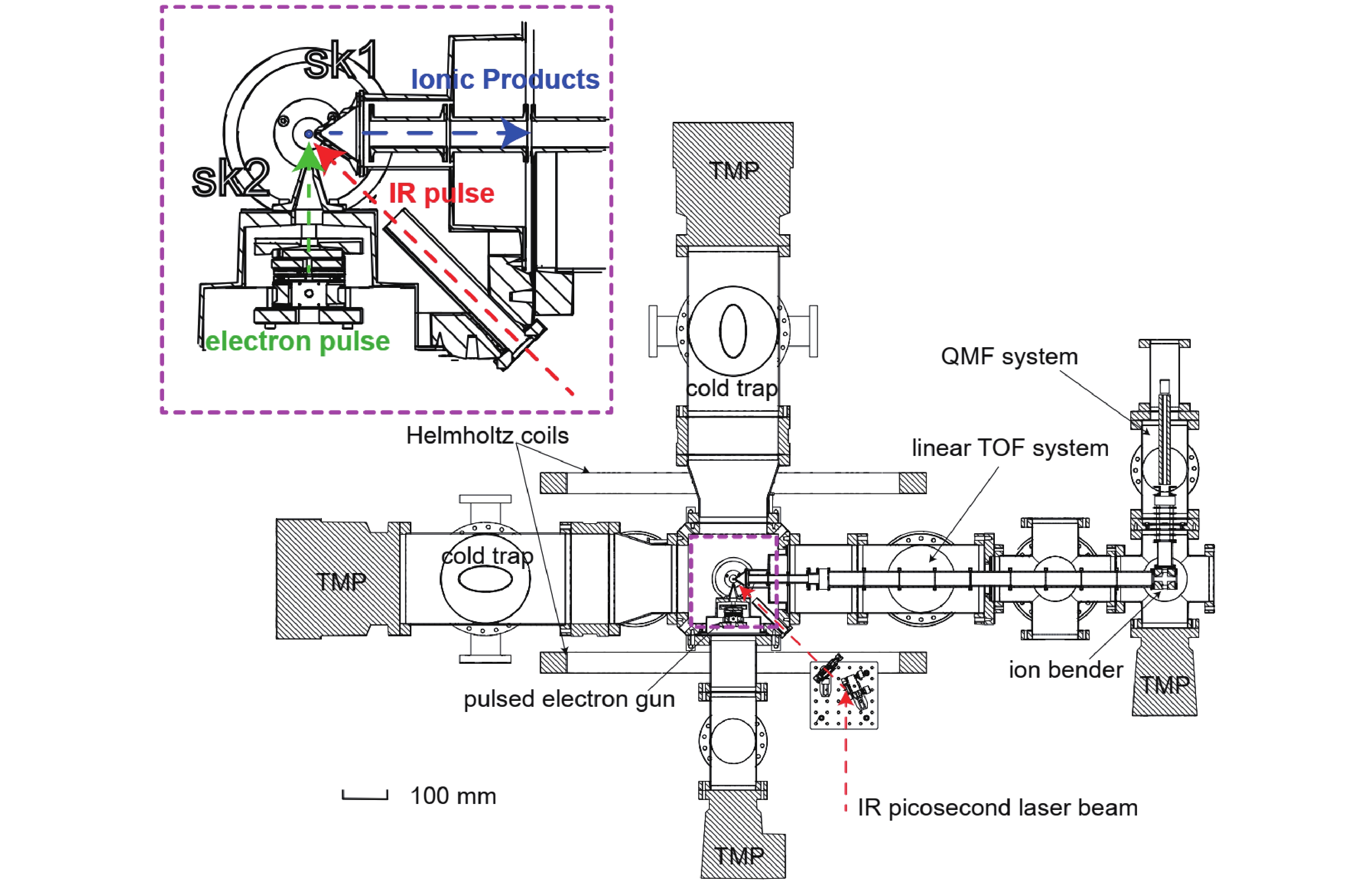
Figure 1. Top view of infrared laser ultra-soft desorption assisted time-delayed mass spectrometer. A liquid beam (diameter ~ 25 μm, noted with a small blue circle in the upper-left zoom-in picture) is perpendicular to the electron-infrared crossed-beam plane. During the experiment, the reaction, electron-gun, and mass-spectrometer chambers are evacuated differentially with two skimmers (SK1 and SK2).
-
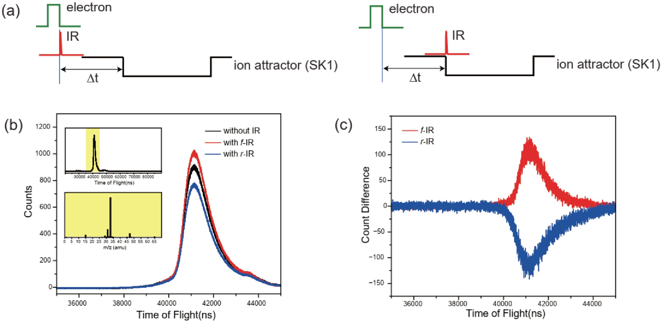
Figure 2. (a) Time sequences, the IR pulse can be set at the rear of electron beam pulse (r-IR, left panel) or in front of ion attracting pulse (f-IR, right panel), (b) IR laser desorption mass spectra, and (c) the differential mass spectra.
-
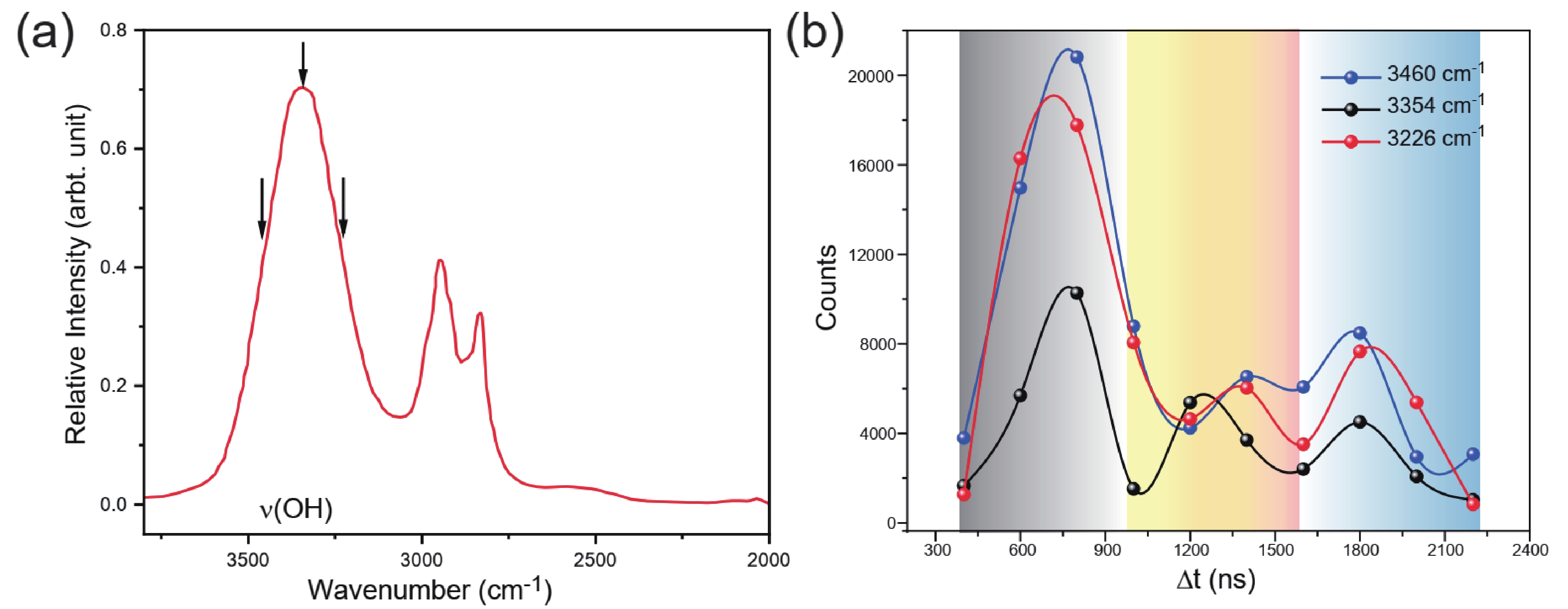
Figure 3. (a) IR laser desorption spectrum of liquid methanol [11] and (b) the enhancements of CH3O+/CH3OH2+ ionic yield observed in the IR desorption assisted time-delayed mass spectra.
Figure
3 ,Table
0 个