2021 Volume 7 Issue 5
Article Contents

Chenyi An, Wei Chen. 2021: Multiplexed single-molecule force spectroscopy for dissecting biophysical regulation of membrane receptors functions on live cells, Biophysics Reports, 7(5): 377-383. doi: 10.52601/bpr.2021.210022
Citation: Chenyi An, Wei Chen. 2021: Multiplexed single-molecule force spectroscopy for dissecting biophysical regulation of membrane receptors functions on live cells, Biophysics Reports, 7(5): 377-383. doi: 10.52601/bpr.2021.210022

Multiplexed single-molecule force spectroscopy for dissecting biophysical regulation of membrane receptors functions on live cells

  • Corresponding author: jackweichen@zju.edu.cn (W. Chen)
  • Received Date: 26/06/2021
    Available Online: 31/10/2021
  • Fund Project: This work was supported by grants from the National Basic Research Program of China (2015CB910800 and 2017ZX10203205), the National Science Foundation of China (31522021 and 31971237), the Fundamental Research Funds for the Central Universities (2015XZZX004-32), and China Postdoctoral Science Foundation (2020M681834).
  • Complex physical cues including two-dimensional membrane environment, dynamic mechanical force, and bioelectric activity inevitably affect membrane receptor functions. Multiplexed single-molecule force spectroscopy (SMFS) techniques with the capability of live-cell measurements are essential to systemically dissect receptor’s functions under complex biophysical regulation. In this review, we summarize recent progress of live-cell based SMFS techniques and specifically focus on the progress of SMFS on the biomembrane force probe with enhanced mechanical stability and multiplexed capability of fluorescence imaging. We further suggest the necessity of developing multiplexed SMFS techniques with simultaneous bioelectric regulation capability to investigate membrane potential regulated membrane receptor functions. These state-of-art multiplexed SMFS techniques will dissect membrane receptors functions in a systematic biophysical angle, resolving the biochemical, biomechanical and bioelectrical regulatory mechanisms in physiologically relevant conditions.
  • 加载中
  • An C, Hu W, Gao J, Ju B-F, Obeidy P, Zhao YC, Tu X, Fang W, Ju LA, Chen W (2020) Ultra-stable biomembrane force probe for accurately determining slow dissociation kinetics of PD-1 blockade antibodies on single living cells. Nano Lett 20: 5133−5140 doi: 10.1021/acs.nanolett.0c01360

    CrossRef Google Scholar Pub Med

    Arlauckas SP, Garris CS, Kohler RH, Kitaoka M, Cuccarese MF, Yang KS, Miller MA, Carlson JC, Freeman GJ, Anthony RM, Weissleder R, Pittet MJ (2017) In vivo imaging reveals a tumor-associated macrophage-mediated resistance pathway in anti-PD-1 therapy. Sci Transl Med 9: eaal3604. https://doi.org/10.1126/scitranslmed.aal3604 doi: 10.1126/scitranslmed.aal3604

    CrossRef Google Scholar Pub Med

    Bashour KT, Gondarenko A, Chen H, Shen K, Liu X, Huse M, Hone JC, Kam LC (2014) CD28 and CD3 have complementary roles in T-cell traction forces. Proc Natl Acad Sci USA 111: 2241−2246 doi: 10.1073/pnas.1315606111

    CrossRef Google Scholar Pub Med

    Brazin KN, Mallis RJ, Boeszoermenyi A, Feng Y, Yoshizawa A, Reche PA, Kaur P, Bi K, Hussey, R E, Duke-Cohan JS, Song L, Wagner G, Arthanari H, Lang MJ, Reinherz EL (2018) The T cell antigen receptor α transmembrane domain coordinates triggering through regulation of bilayer immersion and CD3 subunit associations. Immunity 49: 1−13 doi: 10.1016/j.immuni.2018.06.014

    CrossRef Google Scholar Pub Med

    Brinkmann U, Kontermann RE (2021) Bispecific antibodies. Science 372: 916−917 doi: 10.1126/science.abg1209

    CrossRef Google Scholar Pub Med

    Chang F, Minc N (2014) Electrochemical control of cell and tissue polarity. Annu Rev Cell Dev Bi 30: 317−336 doi: 10.1146/annurev-cellbio-100913-013357

    CrossRef Google Scholar Pub Med

    Chen W, Evans EA, McEver RP, Zhu C (2008) Monitoring receptor-ligand interactions between surfaces by thermal fluctuations. Biophys J 94: 694−701 doi: 10.1529/biophysj.107.117895

    CrossRef Google Scholar Pub Med

    Chen W, Lou J, Evans EA, Zhu C (2012) Observing force-regulated conformational changes and ligand dissociation from a single integrin on cells. J Cell Biol 199: 497−512 doi: 10.1083/jcb.201201091

    CrossRef Google Scholar Pub Med

    Chen W, Lou J, Zhu C (2010) Forcing switch from short- to intermediate- and long-lived states of the αA domain generates LFA-1/ICAM-1 catch bonds. J Biol Chem 285: 35967−35978 doi: 10.1074/jbc.M110.155770

    CrossRef Google Scholar Pub Med

    Chen W, Zhu C (2013) Mechanical regulation of T-cell functions. Immunol Rev 256: 160−176 doi: 10.1111/imr.12122

    CrossRef Google Scholar Pub Med

    De Gast GC, Haagen I-A, Van Houten AA, Klein SC, Duits AJ, De Weger RA, Vroom TM, Clark MR, Phillips J, Van Dijk AJG, De Lau WBM, Bast BJEG (1995) CD8 T cell activation after intravenous administration of CD3×CD19 bispecific antibody in patients with non-Hodgkin lymphoma. Cancer Immunol Immunother 40: 390−396 doi: 10.1007/BF01525390

    CrossRef Google Scholar Pub Med

    De Vlaminck I, Henighan T, Van Loenhout MTJ, Pfeiffer I, Huijts J, Kerssemakers JWK, Katan AJ, Van Langen-Suurling A, Van Der Drift E, Wyman C, Dekker C (2011) Highly parallel magnetic tweezers by targeted DNA tethering. Nano Lett 11: 5489−5493 doi: 10.1021/nl203299e

    CrossRef Google Scholar Pub Med

    Del Rio A, Perez-Jimenez R, Liu R, Roca-Cusachs P, Fernandez JM, Sheets MP (2009) Stretching single talin rod molecules activates vinculin binding. Science 323: 638−641 doi: 10.1126/science.1162912

    CrossRef Google Scholar Pub Med

    Drent E, Poels R, Ruiter R, Van Der Donk NWCJ, Zweegman S, Yuan H, De Bruijn J, Sadelain M, Lokhorst HM, Groen RWJ, Mutis T, Themeli M (2019) Combined CD28 and 4-1BB costimulation potentiates affinity-tuned chimeric antigen receptor-engineered T cells. Clin Cancer Res 25: 4014−4025 doi: 10.1158/1078-0432.CCR-18-2559

    CrossRef Google Scholar Pub Med

    Feng Y, Brazin KN, Kobayashi E, Mallis RJ, Reinherz EL, Lang MJ (2017) Mechanosensing drives acuity of αβ T-cell recognition. Proc Natl Acad Sci USA 114: E8204−E8213 doi: 10.1073/pnas.1703559114

    CrossRef Google Scholar Pub Med

    Ghorashian S, Kramer AM, Onuoha S, Wright G, Bartram J, Richardson R, Albon SJ, Casanovas-Company J, Castro F, Popova B, Villanueva K, Yeung Jenny, Vetharoy W, Guvenel A, Wawrzyniecka PA, Mekkaoui L, Cheung GW-K, Pinner D, Chu J, Lucchini G, Silva J, Ciocarlie O, Lazareva A, Inglott S, Gilmour KC, Ahsan G, Ferrari M, Manzoor S, Champion K, Brooks T, Lopes A, Hackshaw A, Farzaneh F, Chiesa R, Rao K, Bonney D, Samarasinghe S, Coulden N, Vora A, Veys P, Hough R, Wynn R, Pule MA (2019) Enhanced CAR T cell expansion and prolonged persistence in pediatric patients with ALL treated with a low-affinity CD19 CAR. Nat Med 25: 1408−1414 doi: 10.1038/s41591-019-0549-5

    CrossRef Google Scholar Pub Med

    Guo Q, He Y, Lu HP (2015) Interrogating the activities of conformational deformed enzyme by single-molecule fluorescence-magnetic tweezers microscopy. Proc Natl Acad Sci USA 112: 13904−13909 doi: 10.1073/pnas.1506405112

    CrossRef Google Scholar Pub Med

    Hamill OP, Marty A, Neher E, Sakmann B, Sigworth FJ (1981) Improved patch-clamp techniques for high-resolution current recording from cells and cell-free membrane patches. Pflug Arch Eur J Phy 391: 85−100 doi: 10.1007/BF00656997

    CrossRef Google Scholar Pub Med

    He Y, Lu M, Cao J, Lu HP (2012) Manipulating protein conformations by single-molecule AFM-FRET nanoscopy. ACS Nano 6: 1221−1229 doi: 10.1021/nn2038669

    CrossRef Google Scholar Pub Med

    Hong J, Ge C, Jothikumar P, Yuan Z, Liu B, Bai K, Li K, Rittase W, Shinzawa M, Zhang Y, Palin A, Love P, Yu X, Salaita K, Evavold BD, Singer A, Zhu C (2018) A TCR mechanotransduction signaling loop induces negative selection in the thymus. Nat Immunol 19: 1379−1390 doi: 10.1038/s41590-018-0259-z

    CrossRef Google Scholar Pub Med

    Hu KH, Butte MJ (2016) T cell activation requires force generation. J Cell Biol 213: 535−542 doi: 10.1083/jcb.201511053

    CrossRef Google Scholar Pub Med

    Hu W, Zhang Y, Sun X, Zhang T, Xu L, Xie H, Li Z, Liu W, Lou J, Chen W (2019) FcγRIIB-I232T polymorphic change allosterically suppresses ligand binding. Elife 8: e46689. https://doi.org/10.7554/eLife.46689 doi: 10.7554/eLife.46689

    CrossRef Google Scholar Pub Med

    Huang J, Zarnitsyna VI, Liu B, Edwards LJ, Jiang N, Evavold BD, Zhu C (2010) The kinetics of two-dimensional TCR and pMHC interactions determine T-cell responsiveness. Nature 464: 932−936 doi: 10.1038/nature08944

    CrossRef Google Scholar Pub Med

    Johnson KC, Thomas WE (2018) How do we know when single-molecule force spectroscopy really tests single bonds? Biophys J 114: 2032−2039 doi: 10.1016/j.bpj.2018.04.002

    CrossRef Google Scholar Pub Med

    Ju L, Chen Y, Xue L, Du X, Zhu C (2016) Cooperative unfolding of distinctive mechanoreceptor domains transduces force into signals. Elife 5: e15447. https://doi.org/10.7554/eLife.15447 doi: 10.7554/eLife.15447

    CrossRef Google Scholar Pub Med

    June CH, O'Connor RS, Kawalekar OU, Ghassemi S, Milone MC (2018) CAR T cell immunotherapy for human cancer. Science 359: 1361−1365 doi: 10.1126/science.aar6711

    CrossRef Google Scholar Pub Med

    Kim ST, Takeuchi K, Sun Z-YJ, Touma M, Castro CE, Fahmy A, Lang MJ, Wagner G, Reinherz EL (2009) The αβ T cell receptor is an anisotropic mechanosensor. J Biol Chem 284: 31028−31037 doi: 10.1074/jbc.M109.052712

    CrossRef Google Scholar Pub Med

    Kong F, Li Z, Parks WM, Dumbauld DW, Garcia AJ, Mould AP, Humphries MJ, Zhu C (2013) Cyclic mechanical reinforcement of integrin-ligand interactions. Mol Cell 49: 1060−1068 doi: 10.1016/j.molcel.2013.01.015

    CrossRef Google Scholar Pub Med

    Labrijn AF, Janmaat ML, Reichert JM, Parren PWHI (2019) Bispecific antibodies: a mechanistic review of the pipeline. Nat Rev Drug Discov 18: 585−608 doi: 10.1038/s41573-019-0028-1

    CrossRef Google Scholar Pub Med

    Levin M (2021) Bioelectric signaling: reprogrammable circuits underlying embryogenesis, regeneration, and cancer. Cell 184: 1971−1989 doi: 10.1016/j.cell.2021.02.034

    CrossRef Google Scholar Pub Med

    Li R, Ma C, Cai H, Chen W (2020) The CAR T-cell mechanoimmunology at a glance. Adv Sci 7: 2002628. https://doi.org/10.1002/advs.202002628 doi: 10.1002/advs.202002628

    CrossRef Google Scholar Pub Med

    Liu B, Chen W, Evavold BD, Zhu C (2014) Accumulation of dynamic catch bonds between TCR and agonist peptide-MHC triggers T cell signaling. Cell 157: 357−368 doi: 10.1016/j.cell.2014.02.053

    CrossRef Google Scholar Pub Med

    Liu B, Chen W, Natarajan K, Li Z, Margulies DH, Zhu C (2015a) The cellular environment regulates in situ kinetics of T-cell receptor interaction with peptide major histocompatibility complex. Eur J Immunol 45: 2099−2110 doi: 10.1002/eji.201445358

    CrossRef Google Scholar Pub Med

    Liu B, Chen W, Zhu C (2015b) Molecular force spectroscopy on cells. Annu Rev Phys Chem 66: 427−451 doi: 10.1146/annurev-physchem-040214-121742

    CrossRef Google Scholar Pub Med

    Ma L, Cai Y, Li Y, Jiao J, Wu Z, O'Shaughnessy B, Camilli PD, Karatekin E, Zhang Y (2017) Single-molecule force spectroscopy of protein-membrane interactions. Elife 6: e30493. https://doi.org/10.7554/eLife.30493 doi: 10.7554/eLife.30493

    CrossRef Google Scholar Pub Med

    Malmqvist M (1993) Surface plasmon resonance for detection and measurement of antibody-antigen affinity and kinetics. Curr Opin Immunol 5: 282−286 doi: 10.1016/0952-7915(93)90019-O

    CrossRef Google Scholar Pub Med

    Neuman KC, Nagy A (2008) Single-molecule force spectroscopy: optical tweezers, magnetic tweezers and atomic force microscopy. Nat Methods 5: 491−505 doi: 10.1038/nmeth.1218

    CrossRef Google Scholar Pub Med

    Pardoll DM (2012) The blockade of immune checkpoints in cancer immunotherapy. Nat Rev Cancer 12: 252−264 doi: 10.1038/nrc3239

    CrossRef Google Scholar Pub Med

    Porter DL, Levine BL, Kalos M, Bagg A, June CH (2016) Chimeric antigen receptor-modified T cells in chronic lymphoid leukemia. N Engl J Med 365: 725−733

    Google Scholar Pub Med

    Ribeck H, Saleh OA (2008) Multiplexed single-molecule measurements with magnetic tweezers. Rev Sci Instrum 79: 094301. https://doi.org/10.1063/1.2981687 doi: 10.1063/1.2981687

    CrossRef Google Scholar Pub Med

    Rossy J, Laufer JM, Legler DF (2018) Role of mechanotransduction and tension in T cell function. Front Immunol 9: 2638. https://doi.org/10.3389/fimmu.2018.02638 doi: 10.3389/fimmu.2018.02638

    CrossRef Google Scholar Pub Med

    Shi X, Bi Y, Yang W, Guo X, Jiang Y, Wan C, Li L, Bai Y, Guo J, Wang Y, Chen X, Wu B, Sun H, Liu W, Wang J, Xu C (2013) Ca2+ regulates T-cell receptor activation by modulating the charge property of lipids. Nature 493: 111−115 doi: 10.1038/nature11699

    CrossRef Google Scholar Pub Med

    Springer TA, Dustin ML (2012) Integrin inside-out signaling and the immunological synapse. Curr Opin Cell Biol 24: 107−115 doi: 10.1016/j.ceb.2011.10.004

    CrossRef Google Scholar Pub Med

    Struckmeier J, Wahl R, Leuschner M, Nunes J, Janovjak H, Geisler U, Hofmann G, Jähnke T, Daniel JM (2008) Fully automated single-molecule force spectroscopy for screening applications. Nanotechnology 19: 384020. https://doi.org/10.1088/0957-4484/19/38/384020 doi: 10.1088/0957-4484/19/38/384020

    CrossRef Google Scholar Pub Med

    Swamy M, Beck-Garcia K, Beck-Garcia E, Hartl FA, Morath A, Yousefi OS, Dopfer EP, Molnár E, Schulze AK, Blanco R, Borroto A, Martın-Blanco N, Alarcon B, Höfer T, Minguet S, Schamel WWA (2016) A cholesterol-based allostery model of T cell receptor phosphorylation. Immunity 44: 1091−1101 doi: 10.1016/j.immuni.2016.04.011

    CrossRef Google Scholar Pub Med

    Wang X, Ha T (2013) Defining single molecular forces required to activate integrin and notch signaling. Science 340: 991−994 doi: 10.1126/science.1231041

    CrossRef Google Scholar Pub Med

    Wu P, Zhang T, Liu B, Fei P, Cui L, Qin R, Zhu H, Yao D, Martinez RJ, Hu W, An C, Zhang Y, Liu J, Shi J, Fan J, Yin W, Sun J, Zhou C, Zeng X, Xu C, Wang J, Evavold BD, Zhu C, Chen W, Lou J (2019) Mechano-regulation of peptide-MHC class I conformations determines TCR antigen recognition. Mol Cell 73: 1−13 doi: 10.1016/j.molcel.2018.12.012

    CrossRef Google Scholar Pub Med

    Yang M, Brackenbury WJ (2013) Membrane potential and cancer progression. Front Physiol 4: 185. https://doi.org/10.3389/fphys.2013.00185 doi: 10.3389/fphys.2013.00185

    CrossRef Google Scholar Pub Med

    Zhang T, Hu W, Chen W (2021) Plasma membrane integrates biophysical and biochemical regulation to trigger immune receptor functions. Front Immunol 12: 613185. https://doi.org/10.3389/fimmu.2021.613185 doi: 10.3389/fimmu.2021.613185

    CrossRef Google Scholar Pub Med

    Zhou Y, Wong C-O, Cho K-J, Van Der Hoeven D, Liang H, Thakur DP, Luo J, Babic M, Zinsmaier KE, Zhu MX, Hu H, Venkatachalam K, Hancock JF (2015) Membrane potential modulates plasma membrane phospholipid dynamics and K-Ras signaling. Science 349: 873−876 doi: 10.1126/science.aaa5619

    CrossRef Google Scholar Pub Med

    Zhu C, Chen W, Lou J, Rittase W, Li K (2019) Mechanosensing through immunoreceptors. Nat Immunol 10: 1269−1278

    Google Scholar Pub Med

    Zimmerman B, Kelly B, McMillan BJ, Seegar TCM, Dror RO, Kruse AC (2016) Crystal structure of a full-length human tetraspanin reveals a cholesterol-binding pocket. Cell 167: 1041−1051 doi: 10.1016/j.cell.2016.09.056

    CrossRef Google Scholar Pub Med

  • 加载中
通讯作者: 陈斌, bchen63@163.com
  • 1. 

    沈阳化工大学材料科学与工程学院 沈阳 110142

  1. 本站搜索
  2. 百度学术搜索
  3. 万方数据库搜索
  4. CNKI搜索

Figures(3)

Article Metrics

Article views(126) PDF downloads(0) Cited by(0)

Access History

Multiplexed single-molecule force spectroscopy for dissecting biophysical regulation of membrane receptors functions on live cells

Abstract: Complex physical cues including two-dimensional membrane environment, dynamic mechanical force, and bioelectric activity inevitably affect membrane receptor functions. Multiplexed single-molecule force spectroscopy (SMFS) techniques with the capability of live-cell measurements are essential to systemically dissect receptor’s functions under complex biophysical regulation. In this review, we summarize recent progress of live-cell based SMFS techniques and specifically focus on the progress of SMFS on the biomembrane force probe with enhanced mechanical stability and multiplexed capability of fluorescence imaging. We further suggest the necessity of developing multiplexed SMFS techniques with simultaneous bioelectric regulation capability to investigate membrane potential regulated membrane receptor functions. These state-of-art multiplexed SMFS techniques will dissect membrane receptors functions in a systematic biophysical angle, resolving the biochemical, biomechanical and bioelectrical regulatory mechanisms in physiologically relevant conditions.

    • Complex physical cues including two-dimensional membrane environment, dynamic mechanical force, and bioelectric activity inevitably regulate membrane receptor functions. Accurately dissecting their regulatory mechanism is essential to comprehensively understand the working mechanisms of these protein machineries on the membrane. In this regard, single-molecule force spectroscopy (SMFS) techniques arm us powerful tools to characterize membrane receptor’s dynamic functions.

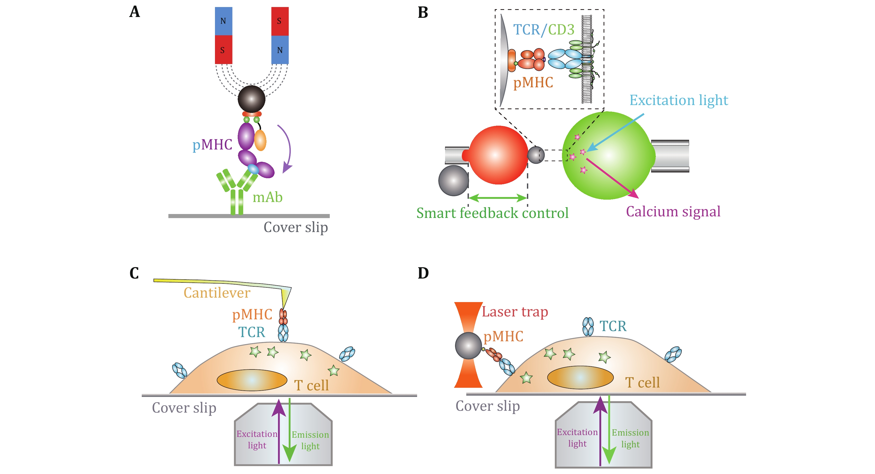
      With the multidisciplinary supports from biophysics and bioengineering fields, it has achieved tremendous progress in the SMFS development and application in biology. SMFS techniques mainly include four platforms, atomic force microscopy (AFM), magnetic tweezers (MT), optical tweezers (OT), and biomembrane force probe (BFP) (Fig. 1). They have revolutionized biological research to reveal the recognition and activation mechanisms of membrane receptors from biophysical standpoint (Chen et al. 2010; Chen and Zhu 2013; Kong et al. 2013; Liu et al. 2014, 2015b; Neuman and Nagy 2008; Wu et al. 2019). In contrast to traditional biochemical ensemble assays, SMFS techniques integrate the advantages of “single-molecule” and “force manipulation”, capable of revealing intermediate conformational or energetic states and binding kinetics of receptor–ligand interactions at the single-molecule level and dissecting receptors’ functions under physiological-relevant forces (Chen et al. 2010; Ju et al. 2016; Liu et al. 2014, 2015a; Zhu et al. 2019). The basic principles, capabilities, and limitations of the four SMFS techniques have been reviewed in detail in (Neuman and Nagy 2008; Liu et al. 2015b). Among the four SMFS techniques, BFP has more appropriate spring constant (0.1–3 pN/nm) for receptor–ligand interactions than AFM (10–105 pN/nm), and more convenient in incorporating live cells without immobilizing cells on substrate. However, the clamping force drifting and the relatively lower degree of automation are still bottlenecks of BFP. In this review, we mainly focus on recent progress of live-cell based SMFS techniques, especially BFP, and their applications in investigating membrane receptor’s dynamics and functions.

      Plasma membrane provides an essential biophysical and biochemical platform to regulate membrane receptor functions. Traditional solution-based biophysical assays (e.g., surface plasmon resonance, SPR (Malmqvist 1993)) and purified protein based SMFS techniques are no longer appropriate to dissect membrane receptor dynamics for the following reasons. First of all, unlike the three-dimensional free diffusion and rotation of proteins in solution, two-dimensional plasma membrane exerts spatial constraint on membrane receptors, thereby limiting receptor’s conformation and spatial location and altering ligand binding kinetics (Huang et al. 2010; Hu et al. 2019). Secondly, the interactions between membrane receptors and plasma membrane (e.g., phospholipids, cholesterol) potentially regulate membrane receptor–ligand recognition (Liu et al. 2015a; Ma et al. 2017; Shi et al. 2013; Swamy et al. 2016; Zhang et al. 2021; Zimmerman et al. 2016). Thirdly, associated complex molecules in cis and downstream signaling cascades can also regulate membrane receptors’ conformations and ligand binding (Chen et al. 2010; Hong et al. 2018; Springer and Dustin 2012). Moreover, biomechanical force from traction forces during cell migration, membrane tension, and cytoskeleton contraction can also regulate membrane receptor’s conformation, ligand binding, and signaling transduction (Bashour et al. 2014; Chen et al. 2012; Hu and Butte 2016; Liu et al. 2014; Wang and Ha 2013; Rossy et al. 2018; Wu et al. 2019). Thus, live-cell based SMFS techniques with multiplexed biophysical manipulation and observation (e.g., mechanical force, fluorescence signal, and bioelectric activity) are required to comprehensively investigate membrane receptor dynamics and functions.

      On the basis of live-cell based SMFS techniques, the development of multiplexed SMFS techniques have enabled direct investigations on mechanisms by which membrane receptor triggers cross-membrane signaling (Feng et al. 2017; Hu and Butte 2016; Ju et al. 2016; Liu et al. 2014) (Fig. 1B1D). Benefiting from its compatibility of live cells and an appropriate force range (1–100 pN) for single-molecule bonds, BFP has advantages in concurrently investigating the ligand recognition and transmembrane signaling at the single-molecule level (Ju et al. 2016; Liu et al. 2014). In the following sections, we mainly focus on BFP-based SMFS techniques and summarize recent technical advances, and suggest the future development of multiplexed SMFS techniques to reveal unprecedent regulatory mechanisms of membrane receptors.

    MECHANICALLY ULTRA-STABLE BFP ENABLES ULTRA-SLOW FORCE-DEPENDENT DISSOCIATION KINETICS MEASUREMENTS ON LIVE CELLS
    • BFP based lived-cell SMFS has demonstrated its power to reveal the mechano-chemistry of weak-transient membrane receptor–ligand interactions, but due to mechanical drifting, characterizing strong receptor–ligand dissociation kinetics (e.g., therapeutic PD-1 antibody dissociating from membrane PD-1) under force on live cells is technically challenging (Chen et al. 2008, 2010), thereby inaccurately estimating ligand binding strength and functions. To overcome this, we recently developed mechanically ultra-stable BFP technique (An et al. 2020) (Fig. 1B). The improvements in clamping force stability and accuracy enable BFP to benchmark the ultra-slow dissociation kinetics of strong interactions between three clinical-approved PD-1 antibodies and PD-1 on live cells at the single-molecule level, revealing the better correlation of the clinical responses of the immunotherapeutic antibodies with their force-dependent dissociation kinetics than commonly accepted solution-based affinity characterized by SPR (An et al. 2020).

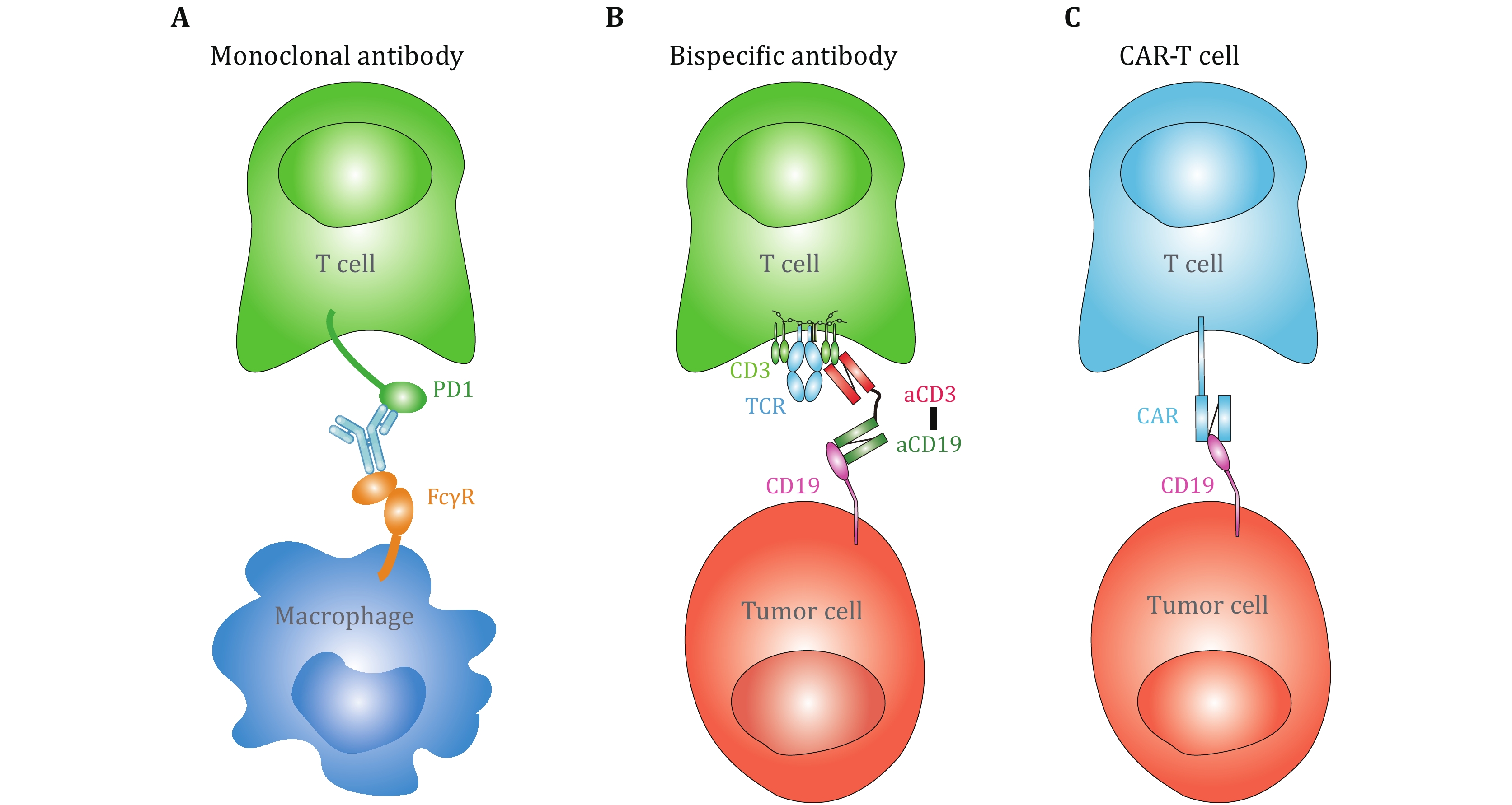
      With this technical advance, live-cell based SMFS measurements may potentially revolutionize immunotherapies. For example, antibody-based immunotherapies, including monoclonal antibodies (mAbs) blocking the ligand binding of immune checkpoint receptors, bispecific antibodies, and chimeric antigen receptor (CAR) T cells, have efficiently boosted cancer therapy (June et al. 2018; Labrijn et al. 2019; Pardoll 2012). One of the bottlenecks impeding accurate prediction of the clinical efficacies of these antibody-based immunotherapies is the inconsistence between the binding kinetics derived from purified proteins based assays (e.g., SPR) and functional/clinical outcomes (An et al. 2020; Drent et al. 2019; Ghorashian et al. 2019). For example, CAT CAR, whose solution-based binding affinity to CD19 is >40-fold lower than that of FMC63 CAR in SPR measurements, has significantly stronger cytotoxicity to CD19-expressing cell line and better clinical outcome (Ghorashian et al. 2019), potentially indicating the requirement of live cell in the kinetic measurements. More importantly, in the following circumstances: (1) potential capture of monoclonal antibodies by Fcγ receptors expressed on myeloid cells (An et al. 2020; Arlauckas et al. 2017) (Fig. 2A), (2) bispecific antibodies physically connect two receptors on opposing cell membranes (de Gast et al. 1995; Brinkmann and Kontermann 2021) (Fig. 2B), and (3) CAR-T recognition of antigenic molecules on target cells (Li et al. 2020; Porter et al. 2016) (Fig. 2C), biomechanical force generated from membrane tension, from cytoskeleton and from cell migration inevitably impose regulatory effects on antibody–antigen binding kinetics. In this consideration, the mechanically ultra-stable BFP technique has excellent potentials in immunotherapeutic applications, such as characterizing the force-regulated antibody–antigen binding kinetics on live cells to provide physiological-relevant biophysical parameters and optimize therapeutic efficacies (An et al. 2020).

    MULTIPLEXED SMFS TECHNIQUES FOR INVESTIGATING MECHANO-CHEMISTRY OF MEMBRANE RECEPTORS
    • Live-cell based SMFS techniques provide the capability to investigate the triggering and transmembrane signaling of membrane receptors. Although SMFS techniques are efficient in revealing ligand recognition process and/or the activation of membrane receptors per se, they are hard to directly observe how membrane receptors react to ligand engagement and transduce the biomechanical stimulations into biochemical signaling cascades inside cells. In this regard, Zhu and colleagues integrated fluorescent imaging with BFP to simultaneously monitor receptor–ligand binding and induced triggering signals (e.g., Ca2+), enabling to uncover the mechanotransduction of membrane receptors resulted from ligand binding under dynamic mechanical force application (Liu et al. 2014). With the fluorescent spectroscopy integrated BFP, Zhu et al. successfully revealed the digital response of force-regulated receptor–ligand binding induced receptor triggering on live T cells (Liu et al. 2014) (Fig. 1B) and platelets (Ju et al. 2016). Nevertheless, multiplexed BFP with higher imaging resolution (e.g., confocal, TIRF) are still demanded. Besides BFP, AFM (He et al. 2012; Hu and Butte 2016), OT (Feng et al. 2017; Kim et al. 2009; Brazin et al. 2018) and MT (del Rio et al. 2009; Guo et al. 2015) have also been integrated with fluorescent spectroscopy successfully. Multiplexed AFM revealed the requirement of biomechanical force in TCR triggered T cell activation (Hu and Butte 2016) (Fig. 1C). Lang et al. determined the direction-dependent biomechanical thresholds for TCR triggering with multiplexed OT (Feng et al. 2017) (Fig. 1D).

    FUTURE TRENDS FOR SMFS TECHNIQUES
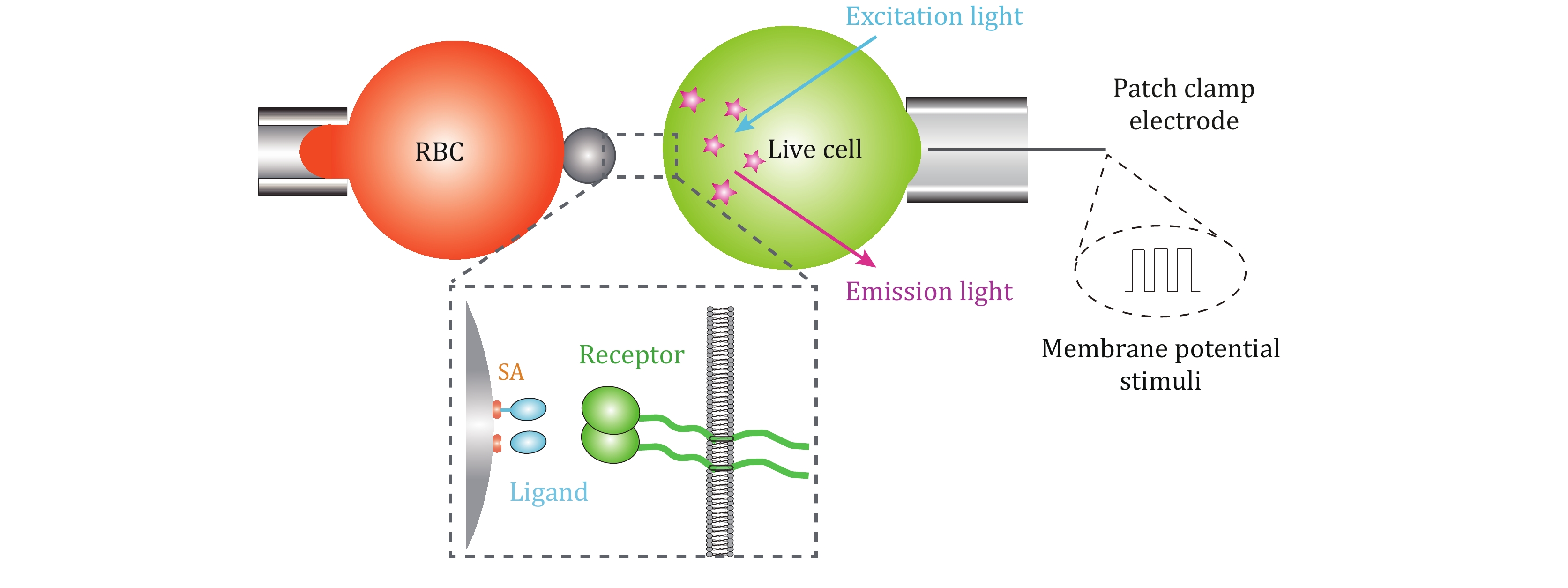
    • Bioelectrical signals are also intrinsic biophysical modulators mediating cellular functions not only in excitable cells but also those non-excitable cells (Yang and Brackenbury 2013). Besides the well-known electrophysiological phenomena in neurobiology, bioelectrical signals have been found to execute crucial modulations in both tumorigenesis and regeneration processes (Chang and Minc 2014; Levin 2021). Although current molecular investigations on bioelectrical mechanisms are mainly confined to ion channels and cytoplasmic cascades (Chang and Minc 2014; Levin 2021), the physiological and pathological alterations of bioelectrical microenvironments potentially regulate the recognition and activation of membrane receptors through mediating the dynamics of receptors, receptor-binding proteins, phospholipids, and ionic concentrations surrounding the plasma membrane (Shi et al. 2013; Zhou et al. 2015). Revealing the interplay of biomechanical, bioelectrical, and biochemical regulatory mechanisms of membrane receptors would further uncover the functional mysteries of membrane receptors and potentially provide new strategies for biomedical applications to conquer cancer and other diseases.

      In order to systematically investigate the complex biophysical regulatory mechanisms of membrane receptors, the multiplexed SMFS techniques require the incorporation of electrophysiological spectroscopy. Upon integrating SMFS techniques with patch clamp, which is efficient in controlling plasma membrane potential (Vm) over the entire cell membrane with its whole-cell mode (Hamill et al. 1981), the biomechanical–bioelectrical coupling regulatory mechanism on membrane receptor–ligand biochemistry would be comprehensively dissected. In addition, further integrating fluorescent imaging technique would uncover membrane receptors’ transmembrane signal transduction modulated by the intersected biomechanical, bioelectrical, and biochemical cues (Fig. 3). In this way, the development of multiplexed SMFS techniques would potentially uncover membrane receptors functions in a systematic biophysical angle and revolutionize relevant biomedical applications.

      Besides, the low efficiencies of SMFS techniques are still bottlenecks hindering their applications in dissecting membrane receptors functions. SMFS experiments require specialized skills and are labor-consuming due to the low-adhesion probability requirement for single-molecule bond (Johnson and Thomas 2018). Although automated AFM and high-throughput MT have partially resolved the time-consuming issues of SMFS techniques (De Vlaminck et al. 2011; Ribeck and Saleh 2008; Struckmeier et al. 2008), improving the maneuverability and throughput of SMFS experiments, especially BFP-based SMFS, is urgently demanded. In this regard, high-throughput tools, such as microfluidic devices, are potential choices to integrate with SMFS techniques in order to enhance their automation and throughput, which would benefit biological discoveries to a larger extent.

    Figure (3)  Reference (52)

Catalog

    /

    DownLoad:  Full-Size Img  PowerPoint
    Return
    Return